Electrical impedance tomography with exploitation of symmetry
View/
Date
2018-01-26Metadata
Show full item recordUsage
This item's downloads: 119 (view details)
Recommended Citation
McDermott, Barry, Porter, Emily, Jones, Marggie, McGinley, Brian, & O'Halloran, Martin. (2018). Electrical impedance tomography with exploitation of symmetry. Paper presented at the Bioengineering in Ireland 2018, 24th Annual Conference of the Section of Bioengineering of the Royal Academy of Medicine in Ireland (BinI 2018), Dublin City University, 26-27 January.
Published Version
Abstract
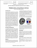
Electrical Impedance Tomography (EIT) is an imaging technique involving an electrode array positioned around the area of interest. Alternating current is injected, and voltage measured, between electrode pairs in a prescribed pattern to produce a measurement set. This set is then processed to generate an image of the area. EIT has several potential biomedical applications including thoracic and neural imaging. Different EIT variants exist with most success being in the production of time difference images. Generation of absolute images (i.e., images generated from a single measurement time) is challenging due to the sensitivity of EIT to errors such as those of electrode placement. This challenge makes imaging of static or very slowly changing scenes, such as those featuring a tumour or an established haemorrhage, very difficult with EIT. In this work, the natural symmetry of an anatomy, such as the sagittal symmetry in the human head, is used to create a difference image without a time change. Measurements are taken from both mirror images of the scene with the difference of the sets processed to produce an image highlighting any deviations in symmetry. This proposed technique allows detection of unilateral pathologies, such as, for example, a bleed in stroke patients.

Except where otherwise noted, this item's license is described as Attribution-NonCommercial-NoDerivs 3.0 Ireland